The marginal position of the placenta. Placenta previa: “hopeless” pregnancy
During pregnancy, the baby is in the placenta. With the help of this shell, the child receives oxygen, nutrients from the mother's body. If the organ is in order and fixed on the posterior wall of the uterus, then the fetus is not in danger of life. A serious pathology during pregnancy is placenta previa syndrome (low or marginal). What danger does the fetus bear, the symptoms of the disease are described below.
What is placenta previa
Improper location or presentation of a child's place is a pathology that is found in the early stages of pregnancy. With this problem, the organ overlaps the internal pharynx partially or completely. It is located in the cervix and can block the birth canal. In the first trimester of pregnancy, pathology is common, but at a later date, “placental migration” may occur - during the development of the baby, the uterus is stretched, the placenta mixes further from the cervix.
Symptoms
The main clinical symptom of placenta previa is bleeding. Its cause is organ detachment: the presence of bloody discharge indicates that part departs from the side walls of the uterus and damages the vessels. Allocate:
- vaginal bleeding;
- internal bleeding (with low previa).
With heavy and frequent bleeding, a woman may suffer from hypotension (low stable pressure) and anemia (hemoglobin level decreases). A pregnant woman is sent to a hospital for storage for constant monitoring and examination. In complex cases, with pathology, fetal death is possible. Bleeding is sudden and always during sleep.

Causes
Placental presentation occurs for many reasons. This can happen after active physical exertion, examination of the cervix by a gynecologist. Pathology can develop in the first weeks. Doctors do nothing until the 24th week: there is a chance of normal movement of the organ and attachment to the walls of the uterus. Other factors include the causes of the appearance of the pathology:
- features characteristic of a fertile egg;
- endometrial pathology;
- cesarean section;
- uterine perforation;
- scraping;
- multiple births with complications;
- myomectomy;
- abnormalities of the location of the uterus;
- uterine contraction;
- diseases of the reproductive system.
Kinds
There are several types of presentation in the cervical region and two main classifications. The first is determined using transvaginal ultrasound diagnostics. The second is determined during childbirth, when the cervix opens 5 cm. The degree and type of pathology changes as the opening of the pharynx, cervix and growth of the uterus changes. There are three options for presentation:
- full;
- low;
- incomplete;
- central;
- lateral.
Full
At full placentation, the placenta overlaps the internal pharynx. That is, if the cervix is \u200b\u200bfully opened, the baby will not be able to be born, because he is blocked by an organ that completely closes the exit from the uterus. With a complete pathology, natural birth is not carried out. One option for delivery is only cesarean section. This location is the most dangerous pathology of the cervix. In 25% of cases during childbirth, serious complications appear that can lead to maternal or child mortality.
Incomplete
In the case of partial presentation (incomplete closure), the organ partially closes the inner channel of the cervix: a small area remains in the opening. Incomplete pathology is compared with a plug, because the organ covers part of the pipe, which does not allow the amniotic fluid to move at the right speed. The lowest edge is flush with the opening of the cervix. The baby’s head cannot pass through the narrow part of the lumen of the birth canal.

Low
The classical low prevalence of chorion during pregnancy is determined by the incorrect location, that is, the organ is 7 cm or more from the perimeter of the cervical canal, does not reach the entrance. The entrance to the area of \u200b\u200bthe internal pharynx of the cervix is \u200b\u200bnot captured. Natural birth can be resolved if the bearing of the fetus proceeds well. Low pathology is the most favorable of all dangerous complications. In obstetric practice, ultrasound determines the degree of pathology during pregnancy.
Central
With this presentation, the entrance to the cervical canal from the uterus is completely closed by a new organ. During a vaginal examination, the gynecologist will not be able to determine the membranes. In this case, there is no natural labor, therefore, a caesarean section is used. Central pathology is determined during childbirth or during a vaginal examination.
Side
During a vaginal examination with lateral presentation, the doctor determines the part of the organ that closes the entrance to the cervical canal, next to which there is a rough fetal membrane. With lateral placentation, an incorrect arrangement is formed, which is determined after examination and corresponds to the results of ultrasound on the presence of incomplete pathology or 2-3 degrees in the first weeks of pregnancy.
Placental presentation
With marginal pathology during a vaginal examination with the help of fingers, the gynecologist is able to determine the rough membranes of the fetus, which protrude into the lumen of the cervical canal. Marginal placentation during pregnancy is determined by the fact that the organ is located near the edge of the internal pharynx. It is determined during a vaginal examination, corresponds to the results of ultrasound by incomplete presentation or 1-2 degrees.
Placenta previa on the back wall
This type of pathology is characterized by the attachment of an organ to the villi of the posterior wall of the uterus. This deviation is frequent with incomplete or low presentation. The main part of the organ is attached to the posterior wall of the uterus, the exit is blocked by the placenta, which prevents natural labor. In this case, a cesarean section is performed - natural childbirth is a danger to the life of the child.
Placenta previa on the anterior wall
Anterior pathology is noted by the attachment of an organ to the anterior wall of the uterus. Such a case is frequent with low or incomplete presentation. That is, the main part of the organ is attached to the front wall of the uterus, while this condition is considered not a pathology, but the norm. This condition is determined during ultrasound up to 26 weeks of pregnancy. At the same time, there is a variant of placental migration, which increases the likelihood that a woman will be sent to a natural normal birth.

What threatens presentation
Placental presentation is periodically repeated, placental abruption can provoke fetal hypoxia and bleeding, therefore, there is a threat of termination of pregnancy. For example, with a complete pathology, it comes to the point that pregnancy ends in premature birth. The consequences of the pathology can be as follows:
- preeclampsia;
- abortion;
- fetoplacental insufficiency;
- incorrect position of the fetus inside the uterus;
- chronic fetal hypoxia;
- leg or pelvic presentation of the fetus;
- iron-deficiency anemia.
Fetoplacental insufficiency is due to the fact that the lower segment of the uterus has a low blood supply, compared with the body or the bottom, that is, there is little blood getting to it. If there is poor blood flow in the localization of the placenta, this means that there is not enough oxygen and nutrients that must flow to the fetus, which does not satisfy its needs. Improper placement of the baby or pelvic presentation due to insufficient space in the lower part of the uterus for the head.
Diagnostics
In order to determine the type or degree of pathology of the placenta, look at the risk factors in history, external uterine bleeding and data from an objective study. An external examination reveals a high standing of the bottom of the uterus (transverse or oblique location of the fetus). Sometimes an auscultation of the noise of the placental vessels in the uterine segment at the location of the placenta is performed. During ultrasound diagnostics are carried out:
- placentation size;
- stage;
- species;
- structure;
- degree of detachment;
- the presence of hematomas;
- threats of abortion;
- migration of the placenta.
During a gynecological examination, an examination of the cervix is \u200b\u200bperformed to exclude vascular injuries or pathologies. With closed external pharynx, part of the fetus cannot be determined. With full presentation, a massive soft formation (fetal bladder) is determined, which occupies the vaginal entrance. During a palpation examination of a pregnant woman, with complete pathology, the occurrence of bleeding is diagnosed. If during the examination in the lumen of the uterine pharynx there are fetal membranes of the uterus and placental tissue, this means that you have an incomplete presentation.
Treatment
Among the methods of treating this pathology, there are two types - drug and non-drug. It is necessary to ensure the complete peace of the woman (exclude physical activity, sex, stressful situations or other). She is prescribed bed rest and drugs, such as Drotaverin, Fenoterol, Dipyridamole, Dexamethasone, which contribute to a better course of childbirth. Caesarean section is prescribed for a narrow pelvis, polyhydramnios, multiple pregnancy, the presence of scars in the uterus.

Birth with placenta previa
With this diagnosis, doctors select an individual approach to delivery. If the mother does not have obstetric complications and other pathologies with low improper attachment of the placenta, this means that there may be a natural birth. During childbirth, the condition of the woman is constantly monitored, especially the amount of spotting that accompanies the process, the indicators of childbirth and the intrauterine condition of the child.
Sometimes urgent tests are performed in the laboratory or ultrasound. If complications, heavy bleeding and full placentation are observed during labor, a cesarean section is performed. Regardless of various complications during pregnancy, it is necessary to act in accordance with the advice of a specialist, so it is recommended to listen to your doctor. Caesarean section at low placentation can also be prescribed.
Prevention
Preventive measures of presentation are the prevention of abortion, the detection and treatment of hormonal dysfunction or genital pathology. Pathology develops during pregnancy and at this time it is necessary to diagnose anomalies. It is recommended to conduct pregnancy rationally, taking into account all threats and risks of complications, timely correct violations in order to obtain optimal delivery.
Video
The placenta is an organ of paramount importance when it comes to pregnancy. Specialists in the field of medicine pay close attention to her during the examination procedure. The placenta is attached to the uterus and grows in parallel with the baby. In appearance, it resembles a kind of flat cake pierced by blood vessels. If the placenta is attached incorrectly or in the wrong place, then such a pathology threatens great difficulties for both the fetus and the expectant mother. The phenomenon can trigger many factors.
Normal placenta
Chorion is converted to the placenta only at the 12th week, but its final maturation occurs only at the sixteenth. After the development of the placenta continues until the 36th week. This body is designed to provide the baby with oxygen, all the necessary substances and trace elements. However, ideal conditions for the normal development of the placenta are not always created.
An interesting fact: according to statistics, about 15% of women experience pathological attachment of the placenta.
 All types of placenta previa are a pathology and require constant monitoring by a doctor
 The physiological norm is the condition when the placenta is attached to the bottom of the uterus or in areas that are close to its lower part: the anterior or posterior wall. If there are deviations, then the body can join the pharynx.
The pharynx is a hole in the uterus that connects it to the vagina. It protects the uterine region from entering the infection.
Based on the location of the placenta, the following types of presentation can be diagnosed:
- complete (the placenta fully covers the uterine pharynx);
- low (the placenta is in close proximity to the pharynx, the approximate distance is 4-5 centimeters);
- lateral (uterine pharynx partially overlaps the placenta);
- marginal (the placenta only touches the pharynx by the edge).
An interesting fact: there is a theory that gravity plays a significant role in deciding where to attach a fetal egg. If the expectant mother prefers to sleep on the right side, then it is attached to the right side of the uterus and vice versa.
What is marginal presentation of the placenta and marginal presentation of the posterior wall
The placenta previa is a pathology that occurs when the upper uterus is unsuitable for implantation of the ovum for a number of reasons, and it is attached below. However, the embryonic organ may “migrate” in the process of bearing the fetus. A change in the location of the placenta occurs due to a change in the structure of the lower uterine segment and due to the lengthening of the upper uterine segment. Usually the process of “migration” begins at the 6th week and ends by the 34th week of pregnancy. In this case, it is not the placenta itself that moves, but the underlying myometrium (the submucous layer of the middle muscle of the uterine wall) shifts. The "migration" of the embryonic organ occurs from the bottom up. If after the 34th week the edge of the placenta still touches the internal pharynx of the uterus, then we can talk about the marginal attachment of the placenta.
An interesting fact: regional placenta previa after 32nd week is characteristic only for 5% of pregnant women. Moreover, they still belong to the risk group, since the percentage of perinatal mortality increases in this case by 25%.
The marginal presentation of the placenta on the posterior wall is an indicator that the organ will not leave the limits of the internal pharynx in most cases. This position will contribute to the successful completion of cesarean section, since the placenta is not injured by an incision. The back wall is not elastic and little susceptible to changes, so the likelihood of "migration" of the embryonic organ is low. Marginal presentation on the front wall is more dangerous, since the organ in this case is subjected to severe stresses, and there is a risk of mechanical damage to the integrity of the placenta. At the same time, there is a high probability that in the later stages of gestation, the placenta will take a normal position.
Regional presentation of the placenta often leads to stable bleeding. The latter are more expected in late pregnancy. This is due to the active formation of the lower uterine segment. The placenta is capable of correctly performing the task assigned to it only when it is located normally.
Important: during pregnancy, it is necessary to control the location of the placenta, its thickness and structure using ultrasound. The first is preferably carried out no later than the 13th week. The thickness of the organ can only be determined at the twentieth.
Complications of regional presentation of the placenta
The placenta can go into a normal position closer to the third trimester. This does not happen only in 5% of women in labor. In this case, the following complications are possible:
- premature labor or the need for emergency termination of pregnancy;
- severe iron deficiency anemia;
- malformations and prolonged hypoxia of the fetus;
- placental abruption (marginal or central);
- rupture of the body of the uterus, due to the fusion of its walls with the placenta;
- perinatal death of the fetus;
- embolism (overlapping gaps) of blood vessels;
- heavy bleeding at the end of labor.
Video: placenta previa
Causes of the pathological location of the placenta
Placenta previa can be caused by a variety of causes and factors. Fetal egg can differ in some features. An important role is played by the state of maternal health and processes occurring directly in the uterus. It will not be possible to affect the place of fixation of the placenta in a medical way, the process is uncontrollable. However, a woman is quite capable of minimizing potential risks.
Anomalies of the ovum
The trophoblast (the outer cell mass of the embryo) that was formed during the cell’s travel through the female reproductive organs is the main assistant at the stage of attachment of the ovum to the uterine wall. In the future, it is he who helps the fetus form the placenta. The membrane covering the fetal egg may be too dense. In this case, successful implantation will not occur, even if the fertilized cell (zygote) is strong.
According to statistics, only healthy embryos, without genetic abnormalities, can correctly invade the uterine cavity. Embryos with congenital abnormalities either do not undergo natural selection by the female body (the latter provokes a miscarriage), or are attached incorrectly.
 Correct implantation of the fetal egg can only occur with good patency of the tubes, the absence of abnormalities in the embryo and the fertile mucous membrane of the uterus
 In addition, the ovum may not be active enough. If it does not release a sufficient amount of enzymes that destroy the mucous membrane in time, then abnormal placentation may occur. While the egg is in the upper segments of the uterus, it does not have time to ripen for implantation, and when the process is completed, then it no longer has a choice and has to be attached lower.
Maternal Health Causes
Once in the uterus, the fetal egg begins to actively search for a place for implantation. Normally, it is attached to the upper layers of the uterus (the posterior wall or the bottom is most often involved). However, this does not happen if the mucous membrane of the organ is damaged. Then the fetal egg descends and penetrates into the lower segments of the uterus. There are a lot of provoking reasons for this phenomenon, their list is as follows:
- addictions;
- inflammatory processes in the uterus;
- frequent birth or a significant number of them;
- carrying out the curettage procedure or abortive intervention during pregnancy, as well as infection, which may be their consequence;
- tumor development in the uterus;
- an abundance of scars on the body of the uterus;
- various abnormalities of the uterine organ;
- endometriosis (a disease associated with the proliferation of internal cells of the uterus outside the body);
- too late first birth;
- hormonal disruptions and disorders;
- multiple pregnancy;
- concomitant diseases of internal organs. With pathologies of the cardiovascular system or circulatory disorders, stagnation in the pelvic organs can form, as a result of which the fetal egg cannot attach normally.
All of the factors described above can negatively affect the course of pregnancy and the development of the fetus.
Symptoms of regional placenta previa
Regional presentation of the placenta can be characterized by two positions of symptoms: mute and pronounced. The first does not involve changes, so a woman is unable to timely and correctly respond to the ongoing process. Violations can only be detected by ultrasound diagnostics.  With an abnormal location, the placenta can break away from the walls of the uterus and provoke bleeding
With a severe form of symptoms, the incorrect location of the embryonic organ is most often manifested by external bleeding. In addition, false contractions may occur at any time. It is the latter that lead to uterine distension, separation of the placenta from its walls and rupture of blood vessels. Bleeding can occur at a time when the organ opens much later than the uterine segment. The placenta exfoliates, which leads to disastrous consequences.
Important: bleeding tends to occur at the most unexpected moment, the process cannot be predicted. It can form even during a night's rest. Its strength and duration also cannot be predicted.
Regional presentation of the placenta can manifest itself in different ways. It all depends on the individual characteristics of the body. At the first sign of discomfort, a doctor’s consultation is required.
Diagnosis of pathological placenta
An anomaly is detected by ultrasound. With the help of ultrasound, it is possible to determine with high accuracy the presence of pathology, the specific position of the body of the placenta and the localization of its edges. Computer diagnostics gives an idea of \u200b\u200bthe thickness of the organ and its size. And also on ultrasound, you can fix the distance from the lower edge of the placenta to the internal pharynx of the uterus. This parameter is very important because it can talk about potential risks and complications.
A bimanual examination of the vagina (assessment of the condition of the uterus, ovaries and pelvic tissues in the gynecological chair) is not advisable to prevent bleeding, which may ultimately cause premature birth. In a situation where it is impossible to conduct an ultrasound, the doctor must carefully examine and draw conclusions.
Treatment
It is impossible to cure the marginal presentation of the placenta in the literal sense of the word. There is only the opportunity to contribute to the “migration” of the embryonic organ or to prevent an aggravation of the situation. In order to reduce pressure on the vessels of the vagina and the lower edge of the placenta, a woman is recommended to use a special bandage. Pregnant in such a situation is contraindicated in physical activity and stress, which can lead to jumps in blood pressure. Sexual contact should also be avoided.  With a diagnosis of placenta previa, a pregnant woman is recommended to wear a bandage
Exercise will help reduce pressure on the lower edge of the placenta: a woman is recommended 3-4 times a day to stand on both hands and legs on the floor. It is necessary to linger in this position for several minutes. Thus, it will be possible to stretch the front wall of the uterus somewhat and achieve some movement of the placenta up. Exercise can be especially effective in the second trimester.  In order to reduce pressure on the lower edge of the placenta, a woman is recommended 3-4 times a day to get on all fours for several minutes
Medication may contain vitamin therapy, taking anti-aggregation (suppressing the adhesion of blood cells) and vascular drugs in doses that are safe for the health of the mother and fetus.
Most often, women with a diagnosis of "marginal placenta previa" are hospitalized for a period of 24 weeks. In the hospital they carry out procedures and preventive measures, such as:
- tocolytic therapy. Pregnant women are prescribed drugs to reduce the number of uterine contractions. This action is possessed by: Ginipral and Partusisten. They are introduced to the expectant mother by the drip or intramuscularly;
- prevention of placental insufficiency. Pregnant women are prescribed vitamin complexes and drugs designed to improve blood circulation: Curantil, Trental or Actovegin;
- prevention of anemia. A woman is prescribed drugs that increase the level of hemoglobin in the blood;
- taking antispasmodics. Women are prescribed candles with papaverine, Magne-B6, No-shpu or magnesium sulfate. Therapy is aimed at reducing the tone of the uterine organ;
- prevention of preterm birth. If there are risks due to placental abruption, additional treatment with corticosteroids: Dexamethasone and Hydrocortisone. This is necessary to prevent respiratory disorders in the infant.
Childbirth with regional presentation
In a situation where special exercises did not help, and the bandage did not give the desired effect, doctors decide on the safest method of delivery. This usually occurs at the 36–38th week of gestation. If the ultrasound still indicates a marginal presentation of the placenta, then the obstetrician-gynecologist may recommend early hospitalization.
If bleeding is mild or absent, then natural delivery is possible. In this case, when the cervix is \u200b\u200bopened in 3 fingers, prophylactic amniotomy is performed (opening the membranes of the membranes).  With the disclosure of the cervix in 3 fingers and the diagnosis of "regional presentation", a prophylactic amniotomy is recommended to a woman
Some obstetrician-gynecologists allow women to give birth on their own, even if bleeding is observed. If the cervix is \u200b\u200bsmooth and soft, then until the contractions an amniotomy is performed, as a result of which the child lowers and is closely pressed to the entrance to the pelvic area, thereby delaying the exfoliated lobes of the placenta. This will stop the bleeding. The woman is also prescribed the drug Oxytocin. It reduces the abundance of blood loss during childbirth and accelerates the process, causing strong and frequent contractions.
When an amniotomy is not effective, a woman with heavy bleeding is prescribed a cesarean section. In some cases, early surgical delivery is acceptable (when the term is less than 36 weeks). In this case, not only a woman, but also a child is prepared for premature intervention by administering drugs, which accelerate the formation of alveoli in the lungs. The maturity of the fetus and its readiness for childbirth will help evaluate ultrasound.
Important: bleeding limits or completely eliminates the use of antiplatelet agents that improve blood flow. Anemia can lead to poor maternal health or fetal hypoxia (lack of oxygen).
Update: October 2018
The placenta previa is considered to be one of the most formidable obstetric pathologies, which is observed in 0.2 - 0.6% of cases of all pregnancies that ended in childbirth. What is the danger of this complication of pregnancy?
First of all, placenta previa is dangerous for bleeding, the intensity and duration of which no doctor can predict. That is why pregnant women with such obstetric pathology belong to the high-risk group and are carefully monitored by doctors.
What does placenta previa mean?
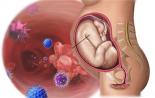
The placenta is a temporary organ and appears only during gestation. With the help of the placenta, the mother and fetus are connected, the child receives nutrients through her blood vessels and gas exchange is carried out. If pregnancy proceeds normally, the placenta is located in the area of \u200b\u200bthe uterine fundus or in the area of \u200b\u200bits walls, usually along the posterior wall, passing to the side walls (in these places the blood supply to the muscle layer is more intense).
The placenta is said to be when the latter is located incorrectly in the uterus, in the region of the lower segment. In fact, placenta previa is when it overlaps the internal pharynx, partially or completely, and is below the pre-existing part of the baby, thus blocking the path for birth.
Types of presentation of the choreon
Several classifications of the described obstetric pathology are known. The following is generally accepted:
Separately, it is worth highlighting low placentation or low placenta previa during pregnancy.
Low placentation - This is the localization of the placenta at the level of 5 or less centimeters from the internal pharynx in the third trimester and at the level of 7 or less centimeters from the internal pharynx during pregnancy up to 26 weeks.
The low location of the placenta is the most favorable option, bleeding during pregnancy and delivery rarely occurs, and the placenta itself is prone to so-called migration, that is, an increase in the distance between it and the internal pharynx. This is due to the stretching of the lower segment at the end of the second and third trimesters and the growth of the placenta in the direction that is better supplied by blood, that is, to the uterine bottom.
In addition, the underlying vessels are isolated. In this case, the vessel / vessels are located in the membranes, which are located in the region of the internal pharynx. This complication poses a threat to the fetus in case of violation of the integrity of the vessel.
Provocative factors
The reasons that cause placenta previa can be related both to the state of the mother's body and to the features of the ovum. The main reason for the development of complications is dystrophic processes in the uterine mucosa. Then the fertilized egg is not able to invade (implant) in the endometrium of the bottom and / or body of the uterus, which forces it to go down. Predisposing factors:

Chronic endometritis, numerous intrauterine manipulations (curettage and abortion), myomatous nodes lead to the formation of an inferior second phase of the endometrium, into which it prepares for implantation of a fertilized egg. Therefore, during the formation of the chorion, she searches for the most favorable place, which is well supplied with blood and optimal for placentation.
The severity of the proteolytic properties of the embryo also plays a role. That is, if the mechanism of the formation of enzymes dissolving the decidual endometrial layer is slowed down, then the egg does not have time to implant in the "necessary" section of the uterus (in the bottom or on the back wall) and goes down below, where it penetrates the mucous membrane.
Symptoms of placenta previa
The course of pregnancy, complicated by placenta previa, is conditionally divided into “mute” and “pronounced” phases. The "mute" phase is almost asymptomatic. During the measurement of the abdomen, the height of the bottom of the uterus is more than normal, due to the high location of the present part of the child. The fetus itself is often located incorrectly in the uterus, there is a high percentage of pelvic, oblique, lateral positions, which is due to the localization of the placenta in the lower part of the uterus (it "forces" the child to take the correct position and presentation).
Symptoms in placenta previa are due to its incorrect localization. The pathognomic sign of this obstetric complication is external bleeding. Hemorrhage from the uterus can occur at any time during pregnancy, but more often in the last weeks of gestation. There are two reasons for this.
- Firstly, in time (Brexton-Gix contractions), which helps to stretch the lower uterus (preparation for childbirth). A placenta, which does not have the ability to contract, “breaks away” from the uterine wall, and bleeding begins from its ruptured vessels.
- Secondly, the “deployment” of the lower segment of the uterus in the second half of pregnancy is intensive, and the placenta does not have time to grow to the appropriate size and it begins to “migrate”, which also causes placental abruption and bleeding.
What is characteristic, bleeding always begins suddenly, often against the background of absolute rest, for example, in a dream. When bleeding occurs and how intense it will be, it is impossible to predict.
Of course, the percentage of profuse bleeding with central presentation is much higher than with incomplete presentation, but this is not necessary. The longer the gestational period, the greater the chance of bleeding.
- For example, marginal presentation of the placenta at a period of 20 weeks may not manifest itself in any way, and bleeding will occur (but not necessarily) only in childbirth.
- Low placentation most often occurs without clinical symptoms, pregnancy and childbirth occur without features.
One of the typical characteristics of bleeding during presentation is their repeatability. That is, every pregnant woman should know about this and always be on her guard.
- The amount of spotting is different: from intense to minor.
- The color of the released blood is always scarlet, and bleeding is painless.
Any minor factor can provoke the occurrence of bleeding:
- straining during bowel movements or during urination
- cough
- intercourse or vaginal examination
Another difference of placenta previa is the progressive anemia of a woman (see). The volume of blood lost almost always does not correspond to the degree of anemia, which is much higher. During repeated bleeding, the blood does not have time to regenerate, its volume remains low, which leads to reduced blood pressure, the development of DIC or hypovolemic shock.
Due to the improper location of the placenta, progressive anemia and a decreased volume of circulating blood, it develops, which leads to intrauterine growth retardation and intrauterine hypoxia.
Case study:In the antenatal clinic, a woman of 35 years was observed - a second, desired pregnancy. At the first ultrasound in a period of 12 weeks, she revealed a central placenta previa. An explanatory conversation was held with the pregnant woman, appropriate recommendations were given, but my colleague, with fear and expectation of bleeding, made an observation. Bleeding for the entire period of pregnancy she had only once, in the period of 28 - 29 weeks, and then, not bleeding, but minor bleeding. Almost the entire pregnancy, the woman was on the sick-list, she was hospitalized in the pathology ward and on threatened dates and during spotting. The woman safely reached almost the deadline and at 36 weeks was sent to the maternity ward, where she successfully prepared for the upcoming planned caesarean section. But, as often happens, on a holiday she began to bleed. Therefore, an operational team was immediately convened. The baby was born wonderful, even without signs). The latter were separated without problems, the uterus contracted well. The postoperative period also proceeded smoothly. Of course, everyone was relieved that such a huge burden fell off his shoulders. But this case is rather atypical of the central presentation, and the woman, one might say, was lucky that everything cost little blood.
How to diagnose?
Placenta previa is a latent and dangerous pathology. If the pregnant woman has not yet had bleeding, then presentation can be suspected, but the diagnosis can only be confirmed using additional examination methods.
A carefully collected history (in the past there were complicated births and / or the postpartum period, numerous abortions, diseases of the uterus and appendages, operations on the uterus, etc.), the course of a real pregnancy (often complicated by the threat of interruption) and external obstetric data helps to suggest a placenta. research.
An external examination measures the height of the uterine fundus, which is longer than the expected gestational age, as well as the incorrect position of the fetus or pelvic presentation. Palpation of the present part does not give a clear sensation, as it hides under the placenta.
In the case of a pregnant woman who complains of bleeding, she is hospitalized in a hospital to exclude or confirm the diagnosis of a similar pathology, where, if possible, an ultrasound scan is performed, preferably with a vaginal probe. Inspection in the mirrors is carried out to establish the source of spotting (from the cervix or varicose veins of the vagina).
The main condition that must be observed when examining the mirrors: the study is carried out against the background of a deployed operating room and necessarily warmed up mirrors, so that in case of increased bleeding, do not slowly proceed with surgery.
Ultrasound remains the safest and most accurate method for determining this pathology. In 98% of cases, the diagnosis is confirmed, false-positive results are observed with an excessively full bladder, so when examining with an ultrasound probe, the bladder should be moderately full.
Ultrasound examination allows not only to establish the presentation of choreon, but to determine its variety, as well as the area of \u200b\u200bthe placenta. The terms of ultrasound during the entire period of gestation are somewhat different from those during normal pregnancy and correspond to 16, 24 - 26 and 34 - 36 weeks.
How pregnant women are led and delivered
With confirmed placenta previa, treatment depends on many circumstances. First of all, the gestational age is taken into account when bleeding occurred, its intensity, the amount of blood loss, the general condition of the pregnant woman and the readiness of the birth canal.
If the presentation of the chorion was established in the first 16 weeks, there is no bloody discharge and the general condition of the woman does not suffer, then she is conducted on an outpatient basis, having previously explained the risks and given the necessary recommendations (sexual rest, limitation of physical activity, prohibition of bathing, visiting baths and saunas).
Upon reaching 24 weeks, the pregnant woman is hospitalized in a hospital where preventive therapy is carried out. Also, all women with bleeding are subject to hospitalization, regardless of its intensity and duration of pregnancy. Treatment of the described obstetric pathology includes:
- medical and protective regimen;
- treatment of fetoplacental insufficiency;
- anemia therapy;
- tocolysis (prevention of uterine contractions).
The medical and protective regimen includes:
- prescription of sedatives (tincture of peony, motherwort or valerian)
- maximum restriction of physical activity (bed rest).
- Therapy of fetoplacental insufficiency prevents the fetal growth retardation and consists in the appointment of:
- antiplatelet agents to improve the rheological qualities of the blood (trental, chimes)
- vitamins (folic acid, vitamins C and E)
- cocarboxylase
- essential Forte and other metabolic preparations
- without fail, iron preparations are indicated to increase hemoglobin (sorbifer-durule s, tardiferon and others).
Tocolytic therapy is carried out not only in the case of a threatened abortion or threatening preterm birth, but also for the purpose of prevention, showing:
- antispasmodics (, magne-B6, sulfate magnesia)
- tocolytics (ginipral, partusisten), which are administered intravenously.
- in case of threatening or beginning preterm birth, respiratory distress is necessarily prevented by corticosteroids and (dexamethasone, hydrocortisone) lasting 2 to 3 days.
If bleeding occurs, the intensity of which threatens the life of a woman, regardless of the gestational age and condition of the fetus (dead or non-viable), abdominal delivery is performed.
What to do and how to deliver when presenting chorion? Doctors raise this question upon reaching the deadline of 37 - 38 weeks. If there is lateral or marginal presentation and there is no bleeding, then in this case the tactics are expectant (the beginning of independent childbirth). When the cervix is \u200b\u200bopened at 3 centimeters, an amniotomy is performed for prophylactic purposes.
If bleeding occurs before regular contractions and the presence of a soft and tensile cervix, an amniotomy is also performed. In this case, the baby’s head lowers and is pressed to the entrance to the small pelvis, and, accordingly, presses the exfoliated lobes of the placenta, which causes the bleeding to stop. If the amniotomy does not produce an effect, the woman is delivered abdominally.
Caesarean section is performed routinely for those pregnant women who are diagnosed with full presentation, or in the presence of incomplete presentation and concomitant pathology (incorrect position of the fetus, pelvic end, age, scar on the uterus, etc.). Moreover, the technique of the operation depends on which wall the placenta is located on. If the placenta is localized along the anterior wall, a corporal caesarean section is performed.
Complications
This obstetric pathology is very often complicated by the threat of interruption, intrauterine hypoxia, and fetal growth retardation. In addition, placenta previa is often accompanied by its true increment. In the third stage of labor and the early postpartum period, the risk of bleeding is high.
Case study:The obstetric department received a multiparous woman with complaints of bleeding for three hours from the birth canal. Diagnosis at admission: Pregnancy 32 weeks. Regional presentation of the placenta. Intrauterine growth retardation of the fetus 2 degrees (by ultrasound). Uterine bleeding. There were no contractions in the woman, the fetal heartbeat was dull, irregular. My colleague and I immediately called the dignity. aviation, since it is still unclear how the matter could end except for the mandatory cesarean section. During the operation was extracted alive. Attempts to remove the placenta were unsuccessful (true increment of the placenta). The scope of the operation was expanded to extirpation of the uterus (the uterus is removed along with the cervix). The woman was transferred to the intensive care unit, where she stayed for a day. The child died on the very first day (prematurity plus intrauterine growth retardation). The woman was left without a uterus and a baby. This is such a sad story, but, thank God, at least they saved the mother.
The slightest changes in well-being during pregnancy are worrying. As a rule, a visit to the doctor should be immediately followed with the hope of hearing that there is no reason to worry and this is a false alarm and the blame for the suspiciousness inherent in all pregnant women. And suddenly it turns out that the fears were not in vain, and the diagnosis “marginal presentation of the placenta” sounds. Instead of starting to panic and drive yourself crazy, you need to calm down, pull yourself together and figure out what it is and how dangerous it is.
The placenta is a unique and complex formation that appears in the woman’s body at the moment when the fertilized egg is attached to the uterine wall. Like any living organism, it goes through all stages of life: appearance, maturation and aging. The life of a small creature that has settled inside her mother’s tummy depends on her. Through it, the baby breathes and receives food. No wonder it is also called a "children's place." It serves as a kind of filter that supplies the fetus with oxygen, and back removes carbon dioxide and metabolic products. Through it, antibodies are transmitted from mother to baby that carry out immune defense. Without it, the same mother’s antibodies would recognize the child as a foreign body and provoke rejection.
Active development of the placenta begins from the 9-10th week. On the 12th child, he completely switches to placental nutrition and receives the official name "fetus". And by the 15-16th week, this is, as a rule, a fully formed organ that will grow with the baby throughout the pregnancy. During planned ultrasounds, they monitor not only the development of the fetus, but also the condition, location and maturity of this vital “cake.”

In the normal course of pregnancy, the placenta is located on the back or front wall of the uterus at a distance from the uterine pharynx. The most optimal and common is considered rear attachment. With him, blood circulation is best, and the place itself is less prone to various injuries. But sometimes it is closer to the exit than it should be, or completely blocks it. This is called a presentation, which, accordingly, can be complete (central) or incomplete.
The most dangerous is full presentation. With him, the birth canal is completely blocked, as a result of which the child can be born exclusively by Caesarean section. 
With incomplete presentation, the placenta is located in the lower segment and partially blocks the exit from the uterus to the cervix. Two types are distinguished: lateral presentation, in which the pharynx overlaps two thirds, and the marginal one, when the lower part of the placenta hangs over the outlet and obscures it no more than a third.
The marginal presentation of the placenta, in turn, happens along the back and front wall, and from the location it has different forecasts:
- On the front wall, on the one hand, is the most dangerous. With it, detachment of the placenta often occurs. The reason for this is that placental tissue is not able to stretch as quickly as uterine tissue. Simply put, it does not have time to grow behind it, and the risk of exfoliation of the hanging edge increases. In addition, this is compounded by the active movements of the child, the physical load of the mother. But, on the other hand, with such regional presentation there is a big chance that with the growth of the uterus, the placenta will rise to a safe distance.
- Along the back wall more common and less threatening than in the previous case. This is due to the fact that this part has less load. With him, there is every chance to calmly endure pregnancy and give birth independently.
In fact, in the world from this pathology, 3-25% of pregnancies end tragically, or the baby is born with some deviations. Therefore, you need to be serious about the regional and another type, regularly monitor the dynamics and follow all the doctor's recommendations.
Causes of regional placenta previa

One of the factors of this pathology is the feature of the fetal egg. After fertilization, the egg descends into the uterus and with its villi attaches to its wall in the upper part. Due to the hormonal background or the structure of the villi, this does not happen. The egg is not able to get to the bottom of the uterus and is attached at the exit.
The main cause of marginal attachment of the placenta is the female body, or rather the condition of the mucous surface or endometrium of the main genital organ.
Factors that violate the integrity of the endometrium and cause presentation, including marginal, are:
- inflammation;
- underdevelopment of the uterus;
- repeated pregnancy;
- endometriosis, endocervicitis;
- genital infections
- age over 35 years;
- scars after abortion or curettage;
- uterine surgery;
- and other benign tumors;
- congenital pathologies;
- diseases of the cardiovascular system;
- diseases of the pelvic organs.
In re-births, this anomaly is observed in 55% of cases, that is, in almost every second. But in general, according to observations, a third of future mothers hear the diagnosis of regional location.

As a rule, they appear at the end of the second or third trimesters from 28 to 32 weeks. At this time, the uterus grows actively. Placental tissue does not have time to stretch and there is an edge detachment, which is accompanied by bleeding. The larger the area that has come off, the more intense they are. This can happen at an earlier stage with or multiple pregnancy.
Any stress can cause detachment. This can happen with intra-abdominal pressure, which is often found in pregnant women, during passionate intercourse and even with a banal raising of the arms. An active baby with his movements can also contribute to this. Often a woman herself serves as a provocateur when lifting weights or with overly active physical exertion. Spotting can also appear while visiting the sauna or taking a hot bath.
Bleeding begins suddenly, without pain, and also suddenly stops. However, it is impossible to predict when this will happen next time and how abundant they will be.

This pathology is sometimes noticed already at the first planned ultrasound at 12-13th week or in the second trimester. As a rule, a woman is not bothered by anything and such a diagnosis sounds very unexpected to her. But most often, the pregnant herself comes to the doctor with complaints of blood discharge or bleeding. After a thorough examination, a conclusion is made about the regional or complete presentation, and the type of abnormal location and the degree of its danger to the fetus and the expectant mother are determined. Given the complexity and risk of complications, in most cases a woman is advised to go to hospital to monitor her condition and complete the examination.
What complications can occur with regional presentation of the placenta?
Due to the fact that when the placenta is detached during the regional location, vessels are damaged, the child does not receive the necessary nutrition and oxygen. This is called fetal hypoxia. She threatens a developmental delay and such consequences for the mother and child as:
- incorrect fetal position;
- —Low hemoglobin — from lack of iron in the body;
- hypotension and, as a result, weakness, and fainting;
- heavy bleeding during childbirth.
- risk of miscarriage;

First of all, it is a complete physical and psychological rest. Not all mothers manage to achieve this at home. Not everyone can afford not to go to work. And it is there that the greatest emotional stress occurs. When an urgent recommendation is made to lie down for preservation, most immediately panic with thoughts about who will work instead of me, as well as washing and cleaning. Believe me, the world will not stop without you. Your main task at the moment is not to submit an accounting report or walk your beloved dog, but to endure and give birth to a healthy, long-awaited baby.
Mom is credited with strict bed rest, an iron-rich diet, and, if necessary, drugs that improve blood circulation, reduce uterine tone, and — increase hemoglobin. Up to 24 weeks, if there is no bleeding and does not bother the general condition, then it is allowed to be treated on an outpatient basis, strictly observing the recommendations and protecting yourself as much as possible from household duties. In difficult situations and at a later date, the mother may be in the clinic until the very birth, and sometimes mainly in the supine position.
The placenta begins to migrate from the end of the second trimester along with a growing tummy. Therefore, after the 26th week there is a good chance that the regional presentation will independently return to normal.

Despite the complexity of the diagnosis, in some cases a woman is still allowed to give birth on her own, but only with a little presentation. The main conditions for this are good labor, a mature cervix and the head location of the fetus. When the neck is opened by more than 4 cm or the finger, the degree of the location of the organ is determined and the amniotic bubble is opened and further childbirth occurs naturally. If bleeding does not stop after opening, then an emergency cesarean is performed. Most often, doctors prefer not to take risks and, with an edge arrangement of any stage, carry out a surgical delivery. This is planned at 38-39 weeks, when the baby is fully formed and ready for the birth.
With a strong or complete presentation
During natural childbirth, there is a high risk of placental detachment, which is fraught with heavy blood loss and other consequences for both the mother and the fetus, up to the death. Therefore, it is better not to take risks and entrust your life and the life of crumbs to experienced professionals.
What precautions should be followed by pregnant women with regional placenta previa

Such a diagnosis is not a sentence, and with it it is quite possible to endure a pregnancy and become a happy mother of a newborn miracle. To do this, you need:
- do not miss scheduled visits to the clinic;
- seek help at the slightest worsening of the condition or the appearance of new symptoms, such as sharp abdominal pain and so on;
- immediately call an ambulance for bleeding;
- just in case, find in advance for yourself several people of their blood type who can become donors for you;
- forget about sex for a while;
- more rest and walk, if it is not prohibited by a doctor;
- sleep for 8 hours;
- follow a diet and do not consume soda and foods that provoke gas formation;
- protect yourself from negative emotions and stressful situations;
- comply with all the recommendations of the attending physician;
- exclude all physical activities: fitness, weight lifting and even cleaning in the house or apartment;
And the most important thing is to enjoy your “interesting position” and to believe that everything will be fine!
Video
Watch the video from which you will find out what presentation is, what other types there are other than marginal, and how it affects the course of pregnancy.
Often, the diagnosis voiced by the doctor serves as an incentive to take a closer look at your health. Especially when you are responsible not only for your life, but also for a small miracle that grows inside you.
Placenta previa is one of the serious complications of pregnancy, in which natural birth is impossible, and in addition, there is a great chance that complications will develop, due to which both the fetus and its mother are at risk of death. In this regard, such pregnancies are carried out with wasp thoroughness, in order to prevent dangerous due to a similar, abnormally located placenta. A woman often has to stay in a hospital for a long time and constantly be under the supervision of doctors. But to make and give birth to a healthy baby is quite possible. It is important to know as much as possible about this pathology.
Placenta previa: what is it?
In the uterus exclusively during pregnancy, with the purpose of nutrition and development of the fetus, a temporary organ is formed - the placenta. It grows and develops in parallel with the fetus, and after its birth, the placenta along with the fetal membranes is separated from the walls of the uterus. From the placenta, which has two sides - the fetal and the maternal, the umbilical cord departs, this is a gelatinous, connective tissue cord, inside of which there are vessels through which blood flows to the fetus, bringing it oxygen and nutrients for life and removing carbon dioxide and metabolic products.

In a physiologically developing pregnancy, the placenta is located in the uterine fundus (the part facing up to the mother’s chest) or along its lateral walls. This is due to the fact that these parts of the uterus are thicker and rich in blood supply, which allows the fetus to develop well and fully, without suffering from a lack of nutrition and oxygen. If, after implantation, the embryo is located in the lower segment, closer to the cervix, and the placenta begins to develop so that during its development it completely or partially blocks the exit from the uterus, they speak of its presentation. This is a complication of pregnancy, which not only violates the nutrition of the fetus, since the walls of the uterus in this part are not so powerful, but also becomes an obstacle to the natural birth of the baby, the exit from the uterus is blocked by the placenta, which can not be born ahead of the baby. If it begins to exfoliate and depart during childbirth, this threatens bleeding and death of the mother and fetus.
Classification of presentation types possible
A similar pregnancy pathology has several classifications, but the following options are most often used:
- Complete presentation , this is the location of the placenta, completely overlapping the internal pharynx of the uterus (the opening through which the baby is born). With this arrangement, the placenta passes in shares from one wall to the second, hanging over the uterine pharynx. At the same time, fetal membranes are not determined during obstetric examination.
- incomplete placenta previa , or partial. With it, the placenta is not completely located above the uterine pharynx, partially overlapping it .

In this type of presentation, two options are distinguished:
- Lateral presentation , with it, one or two lobes of the placenta pass to the uterine pharynx, fetal membranes with their typical roughness are determined.
- Regional presentation , in which the placenta is located in the area of \u200b\u200bthe internal uterine pharynx, but does not leave its limits (that is, the hole is only partially closed).
Low placentation: features
A special option, close to placenta previa, but not so critical, is considered low placentation. With it, there is not even a partial covering of the uterine pharynx from the inside, but the placenta is located critically low to the pharyngeal region, less than 5 cm from the internal pharynx during the third trimester of gestation and less than 70 mm from the internal pharynx for periods up to 26 weeks of gestation.
note
With regard to low placentation during pregnancy, doctors are more supportive in prognoses, especially if it is detected early. With it, bleeding that threatens the life of the fetus and mother rarely occurs, and the placenta, against the background of pregnancy progression, tends to migrate, move up due to the growth of the uterus and stretching of its fibers.
Due to this, as the time increases, it is possible to widen the gap between the uterine pharynx and the edge of the placenta. Usually it rises, due to which the state of low placentation is eliminated by the end of pregnancy, and accordingly, all the complications associated with the presentation and marginal location of the placenta disappear in this condition. With low placentation, childbirth is quite possible in a natural way.
Why is the placenta located incorrectly?
Usually, there must be certain prerequisites for such a complication, and it has been observed that such a pathology is most typical of repeated pregnancies and childbirths. In primiparous, such a complication is rare.
The main factors that can affect the atypical location of the placenta can be changes in the endometrium due to various organic causes (structural problems).
These include inflammatory processes in the uterus as a result of infections, repeated surgical interventions and abortions, operations. Those who left gross scars on the uterus, interventions for the removal of fibroids, as well as after multiple and complicated births. In addition to those listed, it may also matter:
- A large uterus that is not large in the area of \u200b\u200bthe bottom and walls
- , a pathology in which the endometrium is formed in places atypical for it
- Infantilism with underdevelopment of the uterus or malformations in its development (saddle, with septum, bicorn)
- Inflammatory processes in the cervix, anatomical defects from previous births
- The presence of isthmic-cervmcal insufficiency (the cervix is \u200b\u200bweak and cannot hold the fetus).
 The influence of the embryo itself in the period of implantation was also revealed, with the release of special substances. So, when penetrating into the uterus from the fallopian tube, the embryo is implanted in the place where the richest blood network and optimal conditions for development are implanted. During implantation, they secrete special proteolytic enzymes that dissolve the endometrium and immerse it in the thickness. If this process is slowed down, then the embryo cannot attach in the uterine fundus, sliding closer to the cervix, and only there it manages to gain a foothold, subsequently forming a placenta around itself, which will block the exit from the uterus as it grows.
The main symptoms of presentation
note
Actually, before the onset of complications, such a pathology of pregnancy does not manifest itself in any way. It does not give pain, malaise or problems in the growth of the abdomen.
The most frequent and typical manifestation of it, which is also a complication of pregnancy, is recurrent. Bleeding or spotting, daubing can occur at different times, starting from the earliest. About a lot of this is determined by the location of the ovum and the degree of presentation. Most often, repeated bleeding forms from the second half, when the placenta and uterus are actively growing, and the fetus is already pressing on the walls and the placenta. In recent weeks, as training fights become more frequent, there is usually an increase and an increase in the intensity of bleeding.
The main reason for the appearance of blood on the linen during presentation is the detachment of certain sections of the placenta above the uterine pharynx, since the endometrium is capable of active and strong contractions, but the placenta is not. The placenta also does not have time to stretch after the walls of the uterus as pregnancy progresses, and in the zone of greatest stretching and tension, it exfoliates from the wall of the uterus, exposing the vessels that bleed.
Considering the fact that it is the lower segments of the uterus that are most extensible by the end of pregnancy, the fact that the placenta should normally be at the bottom or sides of the uterus is also understandable - there is less contractility and extensibility of tissues.
The presentation may not manifest itself before delivery, but with the beginning of active repetitive contractions of the uterus, the placenta begins to exfoliate, forming life-threatening bleeding and the formation of hematomas between it and the uterine wall, which threatens fetal asphyxiation. At the same time, the woman does not feel any pain, the placenta has no pain receptors, as well as the endometrium, their detachment is invisible to the woman, only blood on the linen and progressive malaise due to blood loss are visible.
note
The blood loses not only the mother, but also the fetus, in addition, through the remaining areas of the attached placenta, it receives little oxygen, which leads to acute and asphyxiation if more than half the area exfoliates.
Features of bleeding: provocateurs and symptoms
Various factors can act as provoking factors for uterine contractions with a similar abnormality of the placenta - physical activity, intimacy, sharp cough tremors against a cold, a doctor examining a chair with an increase in intra-abdominal pressure when straining, taking a hot bath or going to the sauna.
There are some differences in the development of bleeding against the background of full and partial presentation, which should be remembered by the doctor and the most pregnant woman:
If this is a complete presentation:
Bleeding usually occurs against the background of complete health, even without the influence of provoking factors and any subjectively unpleasant, especially painful, sensations. At the same time, it is plentiful and can lead to great blood loss and a threat to life in a short period. It can also spontaneously stop itself, but after a certain period of time it resumes again. Or it may later have the form of discharge of a bloody nature and scanty volume, but appearing regularly. In the last weeks of gestation, bleeding may resume or increase sharply in intensity.
If this is an incomplete presentation:
Bleeding usually occurs at the end of the gestation period, or begins just before the birth, as false contractions intensify or when they transition to true ones. By force, bleeding depends on the volume of the exfoliated area of \u200b\u200bthe placenta, the larger the volume of tissues exfoliated from the uterine wall, the more abundant is the loss of blood. There is no pain or discomfort in the genital tract and lower abdomen.
At the same time, often recurring bleeding, which is typical for presentation, in most cases leads to the development of a post-hemorrhagic form of anemia (occurs from blood loss), in blood tests they give a decrease in hemoglobin and hematocrit in parallel with a decrease in the volume of red blood cells.
What complicates placenta previa
Against the background of previa, pregnancy is often complicated by other pathologies. These include the threat of interruption, which is caused by the same factors as the presentation itself. Often, against the background of presentation, especially if it is complete, are formed, with the birth of a child of varying degrees of prematurity.

In expectant mothers with presentation, hypotension of pregnant women is often detected, it is typical for 35% of cases, which is associated with anemization of the body and a change in vascular tone. In order to avoid all possible complications and bleeding, during the management of pregnant women in the walls of the hospital, they are prescribed drugs to reduce uterine tone and reduce irritability and contractility of the muscles.
Typical for such pregnant women and gestosis is a complication of the second half of pregnancy, in which pressure rises, develops and manifests itself. It leads to sharp violations of the uteroplacental circulation, which leads to hypertension and edema, as well as impaired renal function, due to which protein is massively lost. Presentation both mutually aggravate and aggravate the course of pregnancy.
note
Due to the disruption of the functioning of organs and systems during gestosis, the blood coagulation function is sharply disturbed, which leads to relapse of bleeding, which stops much worse due to the fact that thrombosis does not occur in small vessels.
Also, placenta previa also threatens placenta insufficiency in terms of delivery of oxygen and nutrients to the fetus, which leads to its lag in weight and growth, and leads to the formation of IUGR (baby's underdevelopment syndrome in the womb). That part of the placenta that exfoliates during bleeding is disconnected from functioning, and cannot actively participate in gas exchange and nutrition of the fetus. Back after detachment, this part of the placenta no longer grows, a hematoma is usually formed, which prevents the restoration of the integrity of the placental site.
In the presence of placenta previa, the position of the fetus also suffers, often it becomes transverse or oblique, which is unfavorable for its development and leads to the fact that with its movements the uterus is even more stretched, which creates the preconditions for placental abruption and bleeding. This will not be an obstacle for childbirth, given the fact that they cannot be natural, and with a caesarean section, the position of the fetus will be taken into account by doctors.
How is this anomaly diagnosed?
 Actually, the diagnosis of placental presentation in modern obstetrics has no difficulties. Firstly, the future mother’s complaints about recurring bleeding or spotting, spotting will be typical. Usually repeated from the middle of the gestation period, bloody discharge creates an assumption of complete presentation, that is, an abnormally located placenta on the entire circumference of the uterine pharynx closes the exit. If bleeding occurs in the final weeks of pregnancy or already at the initial stage of childbirth - this is a partial presentation, an abnormally lying placenta only partially blocks the exit.
If bleeding is detected, the doctor must carefully examine the woman on the chair, evaluating the walls of the vagina with the condition of the cervix due to mirrors, in order to exclude traumatic or other causes of bleeding. Also, with a similar vaginal examination of expectant mothers, the signs of an abnormally located placenta are clearly defined. But such techniques can increase bleeding or can provoke other complications.
Therefore, today it is becoming the leading one, and at the same time becoming the most informative in making such a diagnosis. It assesses the location together with the overall size of the placenta, its structure and the detection of bloody clusters between it and the uterine walls. Due to the ultrasound scan, it turns out whether the presentation is complete, or only part of the placental site overlaps the pharynx (which is incomplete). In the study of the placenta, when the state of previa is suspected, it becomes important to determine the placenta area with the exact size of the defect (exfoliated area), the degree of impaired blood flow in the vessels of the fetus and umbilical cord. Also, the dynamics can evaluate the migration of the placenta up to 34 weeks, when it can still change its position due to the growth of the lower segment of the uterus.
Features of ultrasound diagnosis of placenta previa
When a presentation is determined on the basis of data from a study (moreover, it is complete), it is strictly forbidden to use vaginal examinations so as not to provoke bleeding. When there is only partial presentation, its criteria are the length of the gap from the edge of the placenta to the hole that makes up the internal pharynx. If it is 50 mm or smaller, it is a low located placenta. When placental tissue is found in the internal pharynx, this is a presentation. From the period of the second and then third trimesters of gestation, until the period of the 28th week, the position of the placenta is judged by measuring the gap from the inner edges of the uterine throat to the placental tissue, this size is compared with the diameter of the baby’s head.
If an abnormal position of the placenta is detected, it is dynamically controlled according to ultrasound data for up to 34 weeks of pregnancy, when the placenta takes its final position. So, for an assessment in dynamics, at least three studies are carried out at the time of the 16th week, then at 25 weeks and 35 weeks.

It is recommended to conduct an ultrasound with a non-empty bladder, so the position of the uterus and placenta will be better visible. Additionally, due to ultrasound scanning, it is possible to determine the hematoma under the placental site and the uterine wall (if it has already been established that the previa is present). With such a hematoma, bleeding does not always develop and not in all mothers, and there is no outflow of blood from the uterine cavity. If the detachment is not more than 25% of the area, the fetus may not be in danger of life, but if it is increased to one third or more, the fetus may die from
How is pregnancy with a similar problem
 In each situation, the obstetrician-gynecologist will individually select the tactics of pregnancy, based on how often bleeding occurs and how much expressedwhat blood volume is lost by a woman and what are the complications and the degree of anemia. If bleeding did not appear at the beginning of gestation (before the middle of the term), a woman can be at home, being monitored by a doctor at a clinic with a special regime of the day and with the elimination of any provocative factors. Any loads, stresses are limited and a strict ban on intimacy is imposed. Upon reaching the gestational age of 24 weeks or more, in-patient observation is necessary, even if you are feeling fine and there are no spotting and any complaints. Supportive therapy and treatment of all concomitant disorders is carried out in order to bring pregnancy to the prescribed deadlines of 37-38 weeks. This is permissible for rare and mild bleeding, with the normal general condition of the woman herself against the background of a completely normal development of the fetus.
Even if the bleeding was once, and then everything with a pregnancy is good, after a period of 24 weeks she is not discharged from the hospital before the birth of the baby.
If the condition creates fears, then in the conditions of the clinic for pregnant women a special regimen is shown:
- Compliance with bed rest almost always
- the appointment of medicines that relieve the excitability and contractile activity of the myometrium
- Treatment of anemia due to the appointment of iron-containing medications, with severe, not eliminated blood loss - the use of blood-substituting solutions and wildness,
- Correction of uteroplacental insufficiency.
When the pregnancy in the hospital is terminated up to a period of 37 weeks or more, but the state of placenta previa has remained, based on the current situation, delivery is planned and preparation for the operation begins.
How are births done: surgery or not?
 Unconditional indication, carried out as a planned operation, refers to the complete placenta previa. No matter how much the woman would not like, childbirth is physically impossible in a natural way, because the placenta blocks the exit from the uterus, exfoliates before the birth of the fetus and leads to its acute asphyxiation and death in the womb. In addition, the placenta will not allow the present part of the fetus - the head or buttocks, to be inserted into the small pelvis. As the uterus contracts, detachment of the placenta occurs at a progressive pace, from which the mother can die from blood loss during childbirth.
Against the background of incomplete presentation and in the presence of complications in the form of pelvic presentation, improper location of the fetus in the uterus, scarring after surgical labor earlier or with a woman aged 30 years and a narrow pelvis, presence, a cesarean section is also performed as a planned operation. If there are no complications due to incomplete presentation, there is no bleeding and even discharge, the doctor waits for the moment of the beginning of active labor, conducting an amniotomy (opens the fetal bladder). If after this bleeding opens (which means placental abruption), an emergency cesarean section is performed.
If bleeding opens with incomplete presentation before the onset of active labor, a rupture of the fetal bladder is performed. With a similar procedure, the falling head of the fetus in the pelvic area can press the edge of the placenta along the area of \u200b\u200bits exfoliation, and stops bleeding. In a situation where bleeding does not decrease or if there are no signs of cervical maturity, doctors decide - emergency cesarean section . If the neck is mature and there is no more bleeding, preference is given to the delivery of the baby through the natural birth canal.
What complications can pregnant women and women in labor have?
 A similar complication of pregnancy is dangerous both during gestation, and at the beginning of childbirth and after their completion. So, during pregnancy, the threat of interruption and fetal hypoxia, ZVUR syndrome and the death of the child, bleeding are possible. In addition, often with placenta previa, it is also possible to tightly attach it and increment the placenta, which threatens complications in the third period of labor, when the placenta needs to be detached from the uterine walls. If it does not separate, it is necessary to save the uterus for extirpation (removing it with ligation of the vessels) in order to save the mother’s life.
The most common complication is bleeding, which begins both during pregnancy and with the onset of active labor, as labor is more frequent. In this case, doctors resort to partial opening to opening the membranes as an attempt to eliminate the detachment. In order to reduce the risks of such pathologies, doctors in 80% of cases choose for delivery.
Is it possible to give birth to herself with placenta previa
It is naturally possible to give birth at low placentation and partial presentation, if the baby is located head down and the labor is active, the cervix has matured, and there is no bleeding after opening the bladder. In addition to bleeding, complications in such childbirth can be weakness of the labor force and fetal hypoxia developing against this background.
In the management of labor in a natural way, constant CTG monitoring of the fetus and how the uterus contracts is necessary. A woman conducts the first stage of labor with sensors on the abdomen that record uterine activity and contractions, and against them changes in the fetal heart rate. Signs of complications according to monitoring are a reason for immediate emergency caesarean section.
After a natural birth, bleeding may resume due to a violation of the process of separation of the placenta, since it is located in the lower parts of the uterus, which contract worse. It may be necessary to introduce a woman into anesthesia and manual separation of the placenta. Large blood loss may occur due to the decreased tone of the uterus and damage to the vascular plexuses in the neck area with an atypical location of the placenta, which exfoliated over a large area.
Paretskaya Alena, pediatrician, medical observer